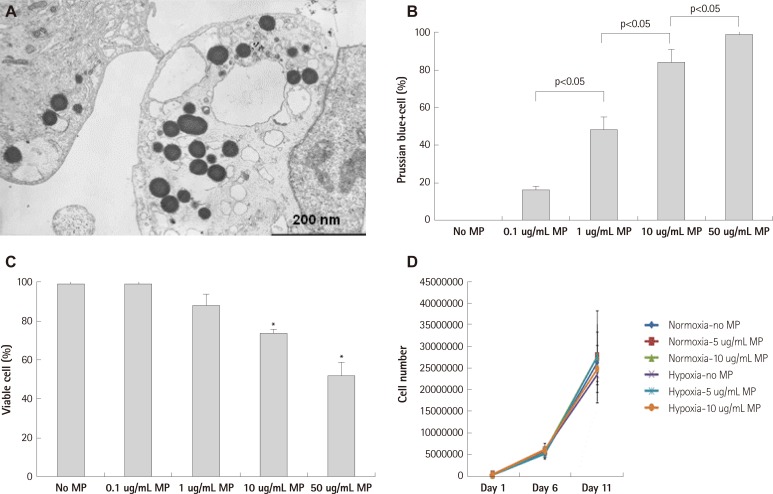
Fig. 1.
Transfection of magnetic bionanoparticle. A: electron microscopic image of endothelial progenitor cell (EPC) which was transfected with magnetic bionanoparticles (MPs). B: transfection efficacy to EPCs was proportionally increased with concentration of MP, which was assessed by Prussian blue staining. C: viability of EPCs was significantly decreased at above 10 ug/mL of MPs, which was assessed by tryphan blue exclusion assay. D: proliferation of EPC was not influenced by MP transfection below threshold concentration (10 ug/mL) in normoxic and hypoxia-reoxygenation condition. There were no significant differences among groups (n=3 respectively). Error bars represented standard deviation. *p<0.05 compared with no MP group (n=3 respectively).

