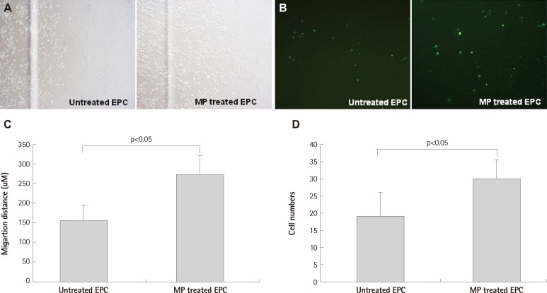
Fig. 3.
Migration of EPC was enhanced by MP transfection with magnet apply. A and C: scratch wound assay: MP was transfected with concentration of 1 ug/mL and magnet was set in cell migrating direction (n=3 respectively). B and D: transendothelial migration assay: magnetic force enhanced MP transfected EPC transmigration through human umbilical vein endothelial cell monolayer: transmigrated cells at bottom of the porous membrane in the vehicle and MPs group (B). EPC were labeled CFSE before co-incubation (n=5 respectively). Error bars represented standard deviation. EPC: endothelial progenitor cell, MP: magnetic bionanoparticle, CFSE: Carboxyfluoroscein Diacetate Succinimidyl Ester.

