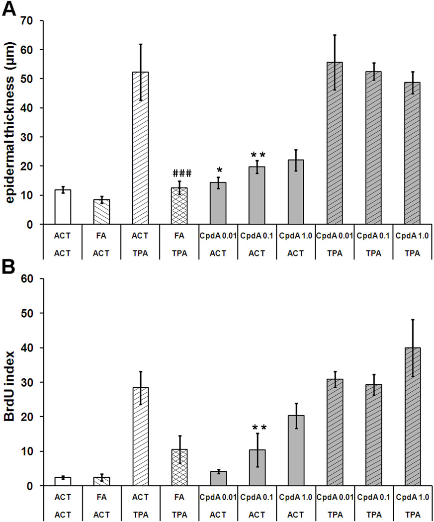
Figure 2. The effect of CpdA on TPA-induced epidermal hyperplasia and cell proliferation in SENCAR mice.
A - the effect of FA (1,5 µg) and CpdA (0.001, 0.1 and 1.0 mg) on the induction of epidermal hyperplasia after multiple treatments with TPA (2 µg). The results of epidermal thickness measurements are plotted as average of twenty measurements at random locations along the epidermis of the skin specimen from each treatment group. B - the effect of tested compounds on BrdU incorporation index in epidermis of SENCAR mice treated with TPA. Proliferative indexes were calculated as the mean percentage of basal layer keratinocytes having BrdU-incorporated nuclei. Approximately 20 randomly selected sites were counted for each sample. The length of skin samples evaluated for the BrdU index as well as for measurement of epidermal thickness was approximately 15 mm Presented results represent the average (±S.D.), P value <0.05(*), <0.01(**), <0.001(***) vs. acetone (ACT) group; P value <0.01 (##), P value <0.001 (###) vs. TPA group.

