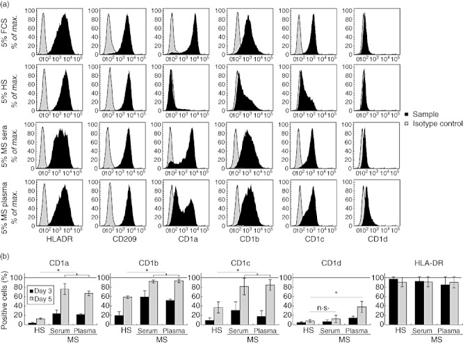
Fig. 1.

Expression of human leucocyte antigen (HLA) class II and CD1 molecules in dendritic cells (DC) differentiated in the presence of sera or plasma from multiple sclerosis (MS) patients. Purified monocytes were cultured in the presence of interleukin (IL)-4 and granulocyte–macrophage colony-stimulating factor (GM-CSF) in medium supplemented with various serum or plasma and were surface-labelled with anti-HLA-DR, -CD209, -CD1a, -CD1b, -CD1c, -CD1d or isotype control fluorochrome-conjugated monoclonal antibodies (mAbs) and analysed by flow cytometry. (a) Profiles of expression in monocytes differentiated in 5% fetal calf serum (FCS), 5% AB human serum [healthy serum (HS)] or 5% MS serum or plasma from one patient (patient 4). Results shown are representative of five independent analyses. (b) Percentage of monocytes cultured in the presence of 5% AB serum (HS) (n = 11), MS serum (n = 11) and MS plasma (n = 9) during 5 days. At days 3 and 5 of differentiation, cells were labelled with anti-CD1a, -CD1b, -CD1c, -CD1d, -HLA-DR or isotype control fluorochrome-conjugated mAbs and analysed by flow cytometry (mean ± standard deviation, Mann–Whitney U-test; n.s.: non-significant; *P-value < 0·05%).
