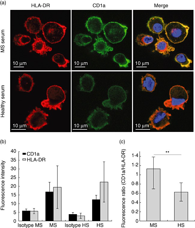
Fig. 2.

Confocal analysis of CD1a and human leucocyte antigen D-related (HLA-DR) expression in monocyte-derived immature dendritic cells (iDC). (a) Images of monocyte-derived DC obtained by confocal microscopy with CD1a in green, HLA-DR in red and nucleus in blue [4',6-diamidino-2-phenylindole (DAPI)], for iDC cultured in patient serum (left) or healthy donor serum (right); (b) fluorescence intensity was quantified in cell images captured by confocal microscopy and averaged as described in Methods. Cells cultured in multiple sclerosis (MS) or in healthy donor (HS) sera were analysed for their expression of CD1a (green channel) and HLA-DR (red channel); (c) normalized expression of CD1a with respect to HLA-DR within each DC analysed in (b) (standard deviation, Student's t-test: n.s.: non-significant, **P-value < 0·01%).
