Fig. 2.

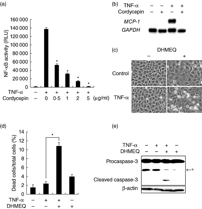
Sensitization to apoptosis by cordycepin through blockade of nuclear factor (NF)-κB. (a) NRK/NF-κB-Luc cells were exposed to tumour necrosis factor (TNF)-α in the presence of serial concentrations of cordycepin for 9 h and subjected to luciferase assay to evaluate NF-κB activity; RLU: relative light unit. (b) Cells were treated with TNF-α in the absence or presence of cordycepin for 6 h and subjected to Northern blot analysis of monocyte chemoattractant protein 1 (MCP-1). The level of glyceraldehyde-3-phosphate dehydrogenase (GAPDH) is shown as a loading control. (c,d) Cells were treated with TNF-α in the absence or presence of 5 µg/ml dehydroxymethylepoxyquinomicin (DHMEQ) and subjected to phase-contrast microscopy (c) and assessment of cell death (d). (e) Cells were treated with indicated agents and subjected to Western blot analysis of caspase-3; *non-specific band.
