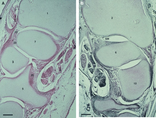
Fig. 6.

(A) Human fetus O.L.-1 (83 mm), week 13. Transverse section (10 μm thick). (1) Distal epiphysis of the radius. (3) Capitate bone. (5) Hamate bone. (7) Lunate. (22) Dorsal radiocarpal ligament. (23) Interosseus ligament. (25) Radiate carpal ligament. Scale bar: 125 μm. (B) Human fetus O.L.-1 (83 mm), week 13. Transverse section (10 μm thick). SM, cell layer which will differentiate into synovial membrane. (2) Distal epiphysis and styloid process of the ulna. (6) Triquetrum bone. (10) Pisiform bone. Scale bar: 125 μm.
