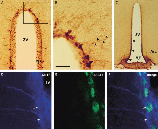
Fig. 4.

P-STAT3 and CNTF IR in the hypothalamus of CNTF-treated adult mice. By peroxidase immunohistochemistry, staining for P-STAT3 is detected in the nuclei of ependymal cells and tanycytes (A). In the latter, the long process is faintly positive (B, arrowheads). The ependymal layer facing the median eminence (ME) and the arcuate nucleus (Arc, arrowheads) exhibits strong P-STAT3 IR (C). Double-label confocal microscopy (D–F) shows a CNTF-positive tanycyte (arrow) also expressing P-STAT3 located near a CNTF-positive tanycyte (arrowhead) not expressing P-STAT3. (B) Enlargement of the framed area in (A). 3V, third ventricle; CNTF, ciliary neurotrophic factor; ep, ependymal layer; P-STAT3, phospho-signal transducer and activator of transcription 3. Scale bars: 80 μm (A); 20 μm (B); 200 μm (C); 10 μm (D–F).
