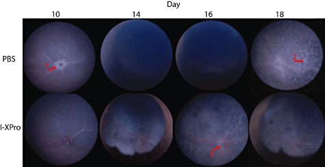
Fig. 2.

Topical endoscopic fundal imaging (TEFI). Experimental autoimmune uveoretinitis (EAU) was induced in B10.RIII mice, and on day 10 post-immunization (p.i.) the mice were treated intraperitoneally (i.p.) with 10 mg/kg [in 100 µl phosphate-buffered saline (PBS)] I-XPro or 100 µ1 PBS. Clinical progression of disease was assessed by TEFI from days 8 to 18; n = 4 mice per group and representative images are shown. In the PBS day 10 image, a raised and swollen optic disc is clearly visible (arrow 1). In the PBS-treated group on days 14 and 16, severe vitritis is causing a vitreous haze, precluding fundal imaging. On day 18 in the PBS-treated group, lesions (arrow 2) that correspond to histological retinal folds are present. In the I-XPro-treated mice on days 14 and 18, development of synechia was noted. On day 16 in the I-XPro-treated group, the optic disc is inflamed (raised and blurred margins; arrow 3) alongside widely distributed perivascular inflammation (vasculitis) and exudative retinal detachment as well as development of inflammatory retinal infiltrates.
