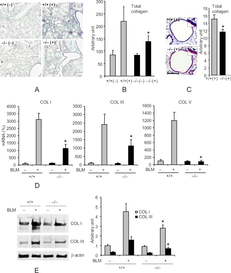
FIGURE 1.
Pin1−/− mice are resistant to BLM-induced lung collagen deposition. A, representative lung sections stained with trichrome for collagen deposition (blue) on day 14 after PBS (−) or BLM (+) challenge. +/+, Pin1 wild type; −/−, knockout. Images are shown at ×10 magnification. B, ImageJ quantification of total collagen staining shown in A. C, left, representative airway stained with trichrome for collagen deposition (blue). Scale bar, 100 μm. Right, ImageJ quantification of total collagen staining. *, p < 0.05 by Student's t test in a two-tailed analysis. No difference was identified between +/+ and −/− mice in base-line collagen levels after PBS treatment. D, qPCR analysis of collagens I, III, and V in lungs of control and BLM-challenged mice on day 14. The mRNA levels in untreated wild-type mice were set as 100% throughout unless otherwise specified. E, immunoblot (left) of collagens in lung of control and BLM-challenged mice on day 14. Right, ImageJ quantification of the immunoblots shown on the left. *, p < 0.05 between BLM-challenged wild type and knockout. No significant differences were identified between +/+ and −/− mice in base-line collagen mRNA and protein levels or after PBS treatment. Data shown are representative of three independent experiments and are expressed as the mean ± S.D. (error bars) of eight animals.

