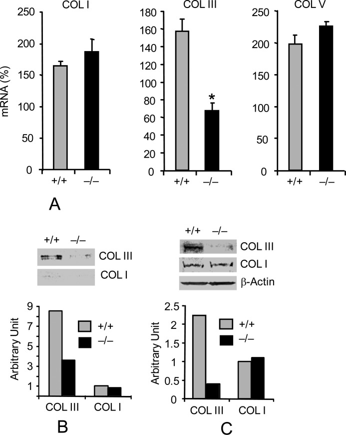
FIGURE 3.
Pin1 knockout decreases type III collagen expression in primary lung Fb. A, wild-type and Pin1−/− primary Fb were starved for 2 days before stimulation with TGF-β1 (1 ng/ml) for 12 h. Total RNA were subjected to qPCR analysis. Data are shown as increased mRNA (percentage) after TGF-β1 compared with the respective untreated control. Error bars, S.D. of 3–4 separate cultures in each group. Data are representative of at least three independent experiments. *, p < 0.05 by Student's t test in a two-tailed analysis. B, cells were treated as in A, and secreted collagens in the culture medium were immunoblotted (top) with anti-collagen I and III. ImageJ quantification (bottom) of the immunoblots from two independent experiments. C, cell lysates were immunoblotted (top) with anti-collagen antibodies shown. ImageJ quantification (bottom) of the immunoblots from two independent experiments.

