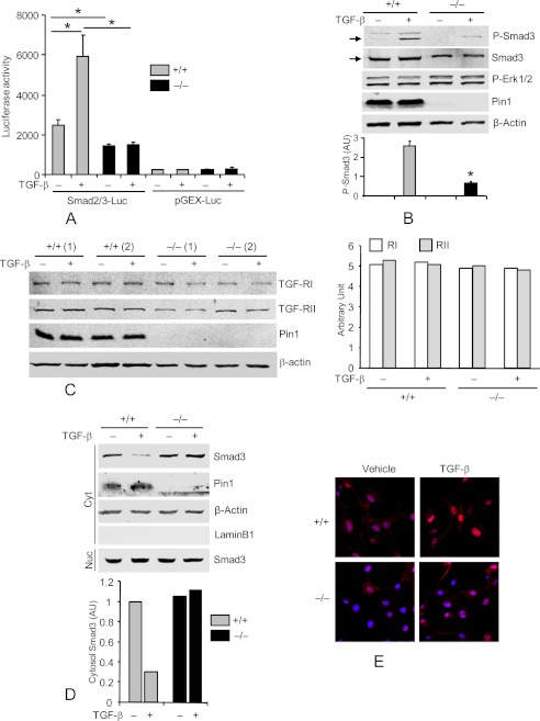
FIGURE 5.
Pin knockout suppresses TGF-β1-Smad signaling in primary lung Fb. A, cells were infected with lentiviral vectors containing Smad2/3-responsive luciferase reporter and Renilla-luciferase constitutive reporter. After 2 days, cultures were treated with vehicle or TGF-β1 (1 ng/ml) for an additional 2 days. Cell lysates were subjected to luciferase activity assay using the Dual-Luciferase Assay system and normalized to the Renilla-luciferase reporter. Lenti-pGEX-luc without Smad2/3 binding sites served as negative control. B, cells were starved for 2 days and treated with TGF-β1 for 1 h. Cell lysates were immunoblotted with antibodies to phospho-Smad3 (Ser534/536), total Smad3, phospho-Erk1/2, Pin1, and β-actin. The ratios of Smad3 phosphorylation to the total Smad3 were quantified by ImageJ (bottom). AU, arbitrary units. C, cells from two mice were treated as in B, and total cell lysates were immunoblotted with anti-type I and II TGF-β1 receptors. ImageJ quantification of the immunoblots is shown on the right. D, cells were treated as in B, and cytoplasmic (Cyt) and nuclear (Nuc) extracts were immunoblotted (top) with antibodies to total Smad3, Pin1, β-actin, and lamin B1 (nuclear marker) (Nuc). C+, positive control. Bottom, ImageJ quantification of the immunoblots from two independent experiments. E, cells were treated as in B and immunostained with anti-Smad3 (red) and TO-Pro (nuclear dye; blue). *, p < 0.05. Data shown are representative of five independent experiments and are expressed as the mean ± S.D.

