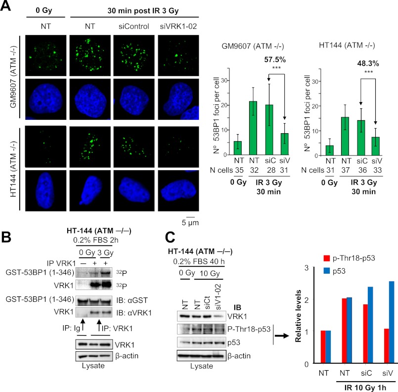
FIGURE 4.
Defective 53BP1 foci in ATM−/− cell lines by loss of VRK1. A, HT144 and GM9607 cells were transfected with siVRK1-02 or siControl or not transfected (NT). After 72 h, cells were either untreated or exposed to 3 Gy of IR and allowed to recover for 30 min. Cells were immunostained with 53BP1 polyclonal antibody. The quantification of the number of 53BP1 foci per cell in each condition is shown in the graphs. Means and S.D. are represented. The number of cells analyzed is indicated below. Images showing several cells and VRK1 protein levels detected by Western blot are shown in supplemental Fig. S5. ***, p < 0.001. B, activation of VRK1 in response to IR in HT144 cells. Endogenous VRK1 was immunoprecipitated from non-irradiated or 3-Gy irradiated HT144 starved-cells. This immunoprecipitate was used to assess VRK1 autophosphorylation and phosphorylation of GST-53BP1(1–346) in an in vitro kinase assay. Ig is a negative control with a nonspecific antibody (anti-HA). C, phosphorylation of p53 at Thr-18 is dependent on VRK1 in HT144 cells. HT144 cells were transfected with siVRK1-02 or siControl or not transfected (NT). After 24 h, cells were serum-starved for 40 h and then irradiated with 10 Gy of IR or left untreated. Cell lysates were prepared 1 h after irradiation, and Western blots were performed with antibodies to VRK1 (1B5), phospho-Thr-18-p53, p53, and actin. Quantification of the levels of phospho-Thr-18-p53 and total p53 is shown in the graph. IP, immunoprecipitation; IB, immunoblot. Error bars represent the standard deviation.

