FIGURE 6.
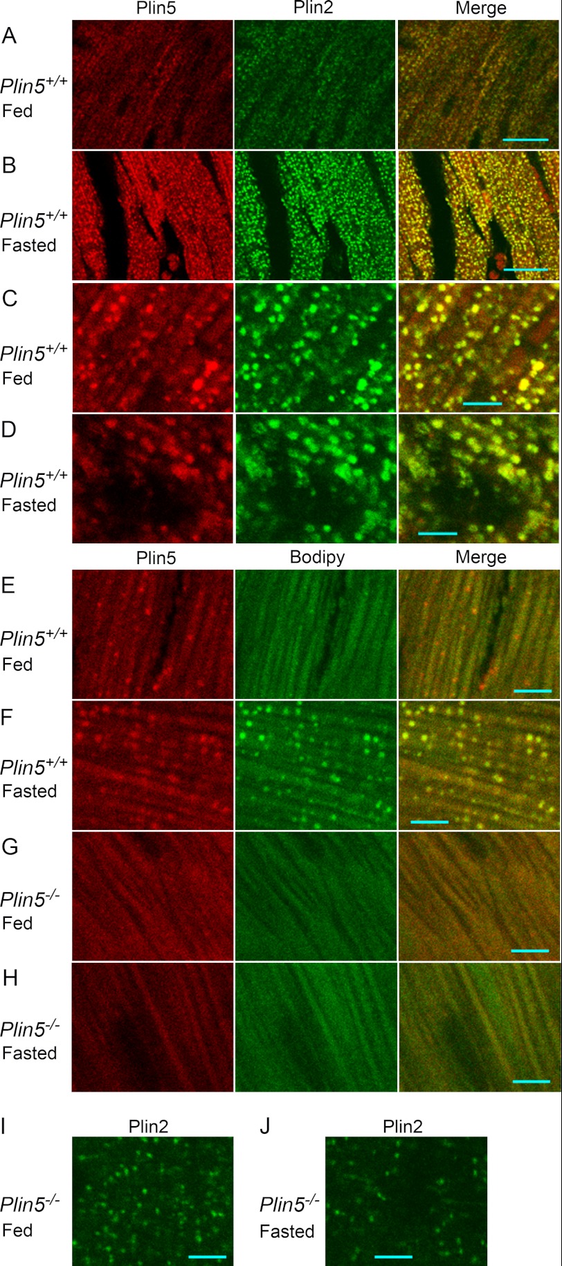
Immunofluorescence staining of heart sections. A–D, immunofluorescence staining of Plin5 and Plin2 in heart sections from wild-type mice. A and C, fed mice; and B and D, starved mice. A and B, low magnification. Bar, 20 μm. C and D, high magnification. Bar, 5 μm. E–H, double staining with an anti-Plin5 antibody and Bodipy 493/503. Cryosections of the hearts of wild-type (E and F) and Plin5−/− (G and H) mice under fed (E and G) and fasted (F and H) conditions were stained. I and J, immunostaining of Plin2 in Plin5−/− mice under fed (I) and fasted (J) conditions. Bar, 5 μm.

