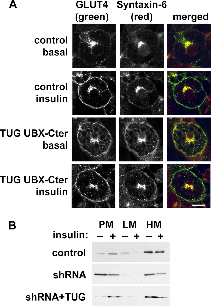
FIGURE 6.
TUG disruption mobilizes syntaxin-6 to the plasma membrane. A, basal and insulin-treated control and UBX-Cter-containing 3T3-L1 adipocytes were imaged using confocal microscopy. GLUT4 was detected by a GFP tag, and syntaxin-6 was detected by immunostaining. Scale bar, 10 μm. B, control, shRNA, and shRNA + TUG 3T3-L1 adipocytes were treated with insulin, subjected to subcellular fractionation, and immunoblotted as indicated to detect syntaxin-6. Experiments were repeated at least twice with similar results.

