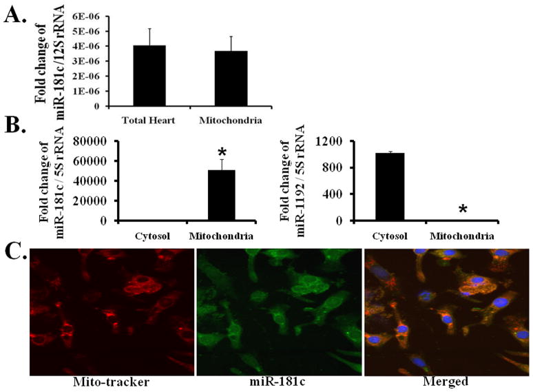
Figure 2. Mitochondrial localization of miR-181c.
qRT-PCR shows that miR-181c is mainly present in the mitochondria.
(A) miR-181c expression is almost the same in total RNA derived from the mitochondrial fraction and the total heart fraction. 12S rRNA is the internal control to normalize these data.
(B) miR-181c expression is mainly detected in the mitochondrial fraction and not in the cytosolic fraction (left panel), whereas miR-1191 is mainly present in the cytosolic fraction (right panel). 5S rRNA is the internal control to normalize these data, as this RNA is present in both cytosol and mitochondrial fractions. *p<0.05 vs. Cytosol.
(C) Fluorescent in situ hybridization demonstrating intramitochondrial localization of miR-181c. Mitochondria are labeled with mito-tracker red (left), miR-181c is green (GFP, middle), and the merged image is shown in the right panel.

