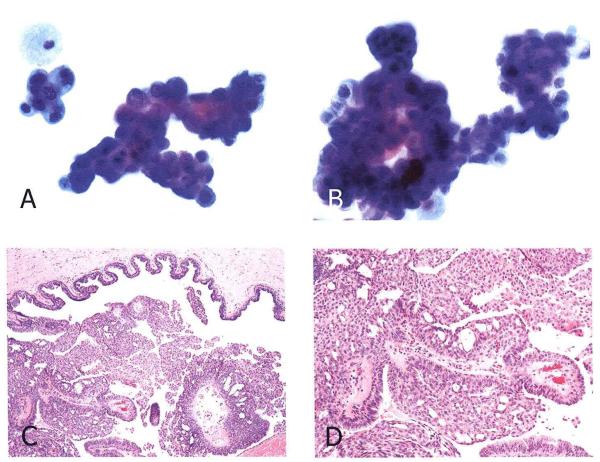
Figure 2.
False positive cytology in a case of intraductal papilloma with atypia. (A) Two clusters of epithelial cells exhibiting nuclear enlargement, hyperchromasia and distinct nucleoli. A mitotic figure is evident in the center of left upper cluster. This NAF specimen was called suspicious for malignancy. (B) MD cytology from the same patient which was independently called suspicious for carcinoma. A large, branching papillary cluster of epithelial cells exhibits marked nuclear enlargement and hyperchromasia. (Papanicolaou stain, original magnification × 500) (C, D) histologic section of the excisional biopsy exhibiting an intraductal papilloma with areas of atypical hyperplasia (H & E stain, original magnifications C, ×50; D, ×100)

