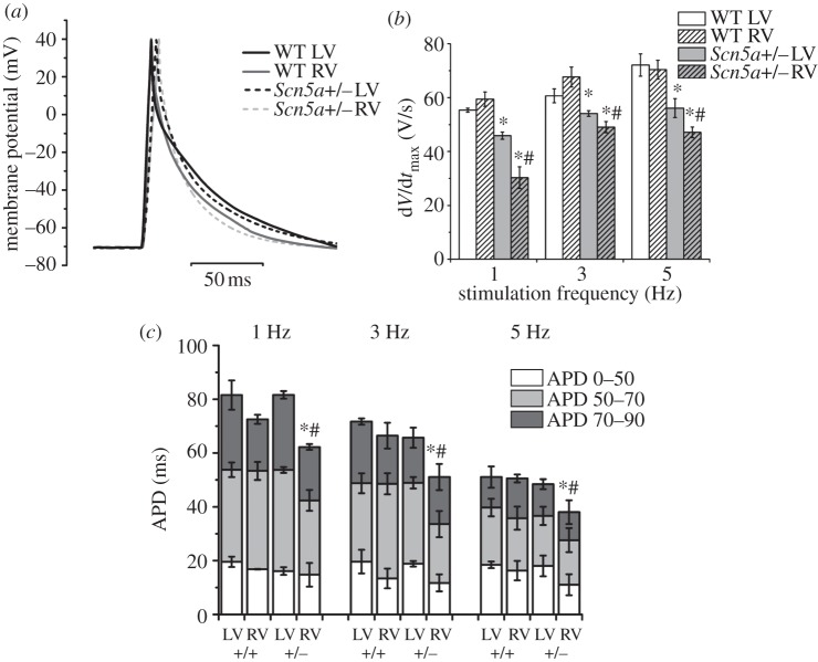
Figure 3.
AP measurements. (a) Examples of AP traces at 1 Hz pacing overlaid from the LV and RV of WT and Scn5a+/− myocytes. (b) Bar chart comparing upstroke velocity. (c) Bar chart comparing APDs with APD50s, APD70s and APD90s superimposed. Significant differences: asterisks (*), effect of genotype; hashes (#), effect of cardiac ventricle (n = 20 for each group).

