Figure 1.
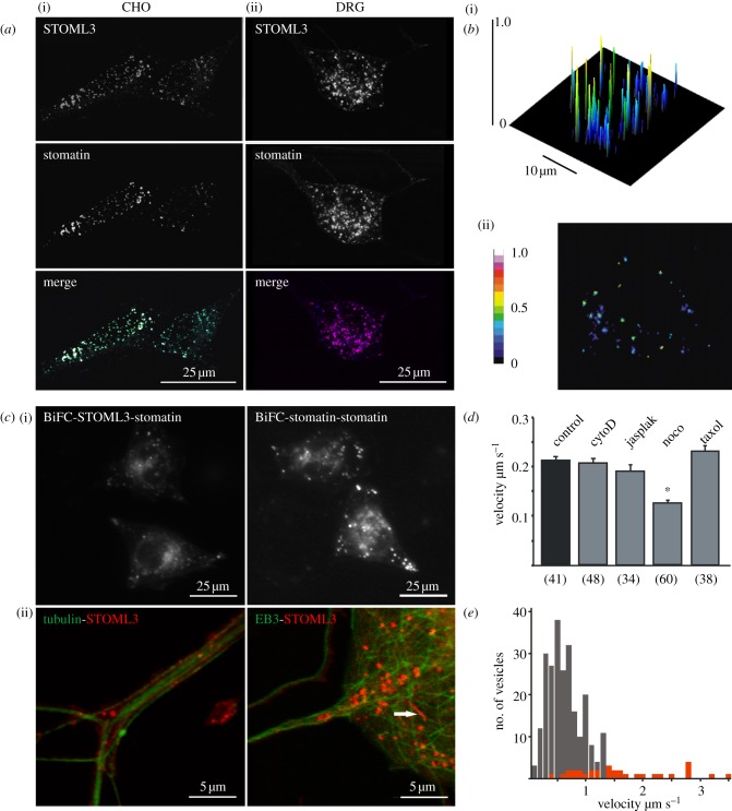
STOML3 and stomatin localize to microtubule-associated vesicles (a) STOML3 co-localizes with stomatin in CHO cells (i) and DRG neurons (ii). (b) Representative image of FRET efficiency in a single CHO cell co-expressing STOML3-AmCyan1 and EYFP-stomatin. A two-dimensional image (ii) and surface plot with the FRET efficiency projected in colour onto the z-axis (i) are shown. (c) In CHO cells, stomatin interacts with STOML3 and stomatin itself, as demonstrated using BiFC (i). (ii) Also shown is the co-localization of STOML3 (red) with EB3 or tubulin (green) in DRG neurons. Note that some STOML3 vesicles are very long (arrow). (d) Quantification of STOML3-particle motility in CHO cells treated with compounds that destabilize or stabilize actin filaments (cytoD, cytochalasinD; jasplak, jasplakinolide) or microtubules (noco, nocodazole; taxol), respectively. *p < 0.05 versus untreated control. Numbers in parentheses indicate number of cells. (e) Frequency distribution of STOML3-positive particle velocities measured for all moving puncta (grey bars) or only for vesicles more than 1 µm in length (red bars). Note that longer vesicles travel with much higher average velocity than the non-selected population.

