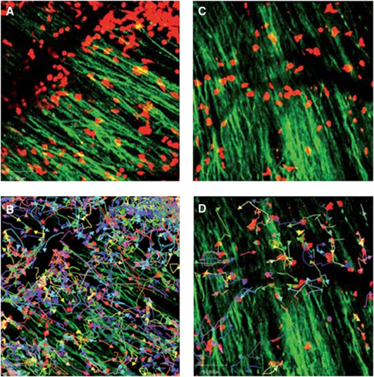
Figure 5.
Immune infiltrates in demyelinating lesions as depicted by two-photon laser scanning microscopy (TPLSM) are highly dynamic and show different motility patterns in distinct disease stages. (A) TPLSM of a representative brainstem lesion in the onset of EAE in B6.tdRFP/B6.Thy1.EGFP (green: EGFP, neuronal processes; red: CD45+.tdRFP cells) (maximal intensity projection of a volume of 70 mm thickness and 36 planes, time point 0). (B) Automated single-cell tracking of CD45+.tdRFP cells. (C) TPLSM of a representative brainstem lesion in the onset of 2d2.tdRFP Th17 cells (red)-induced passive EAE in B6.Rag1/Thy1.EGFP. (D) Automated single-cell tracking of 2d2.tdRFP Th17 cells. (Figure reprinted with permission from Siffrin et al, 2010).

