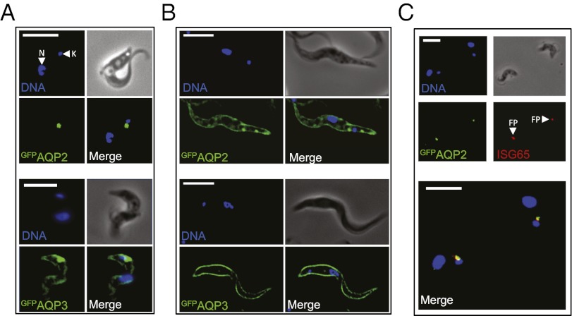
Fig. 3.
AQP2 is restricted to the flagellar pocket specifically in bloodstream-form cells. (A) Fluorescence microscopy reveals the location of GFPAQP2 and GFPAQP3 in bloodstream-form cells. DNA is stained with DAPI and the nucleus (N) and mitochondrial genome (kinetoplast, K) are indicated. (B) Fluorescence microscopy reveals the location of GFPAQP2 and GFPAQP3 in insect-stage cells. (C) Immunofluorescence microscopy reveals the location of GFPAQP2 colocalized with the flagellar pocket (FP) marker, ISG65 in bloodstream-form cells. GFPAQPs are expressed in an aqp2/aqp3 null background. (Scale bars, 10 μm.)

