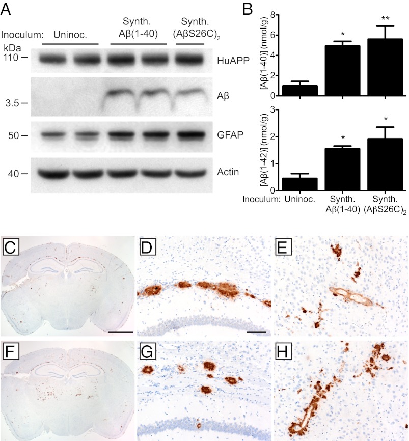
Fig. 4.
Induction of cerebral Aβ deposition in Tg(APP23:Gfap-luc) mice inoculated with synthetic Aβ aggregates. (A) Immunoblotting shows that increased GFAP and Aβ protein levels were apparent in brain homogenates prepared from male Tg(APP23:Gfap-luc) mice inoculated with synthetic Aβ aggregates at 330 dpi compared with age-matched, uninoculated Tg(APP23) mice. Actin levels are shown as a control. (B) By ELISA, Aβ(1–40) (Upper) and Aβ(1–42) (Lower) levels were significantly increased in Tg(APP23:Gfap-luc) mice inoculated with synthetic Aβ(1–40) or (AβS26C)2 aggregates at 330 dpi compared with age-matched, uninoculated controls (n = 4 each). *P < 0.05, **P < 0.01. Data are mean ± SEM (C–H) Aβ immunostaining of brain sections from female Tg(APP23:Gfap-luc) mice inoculated with synthetic Aβ(1–40) (C–E) or (AβS26C)2 (F–H) aggregates at 330 dpi. Induced Aβ deposition was apparent in whole brain coronal sections (C and F) as well as the corpus callosum (D and G) and thalamus (E and H) of inoculated mice. (Scale bar in C, 2 mm and also applies to F; scale bar in D, 100 μm and also applies to E, G, and H.)

