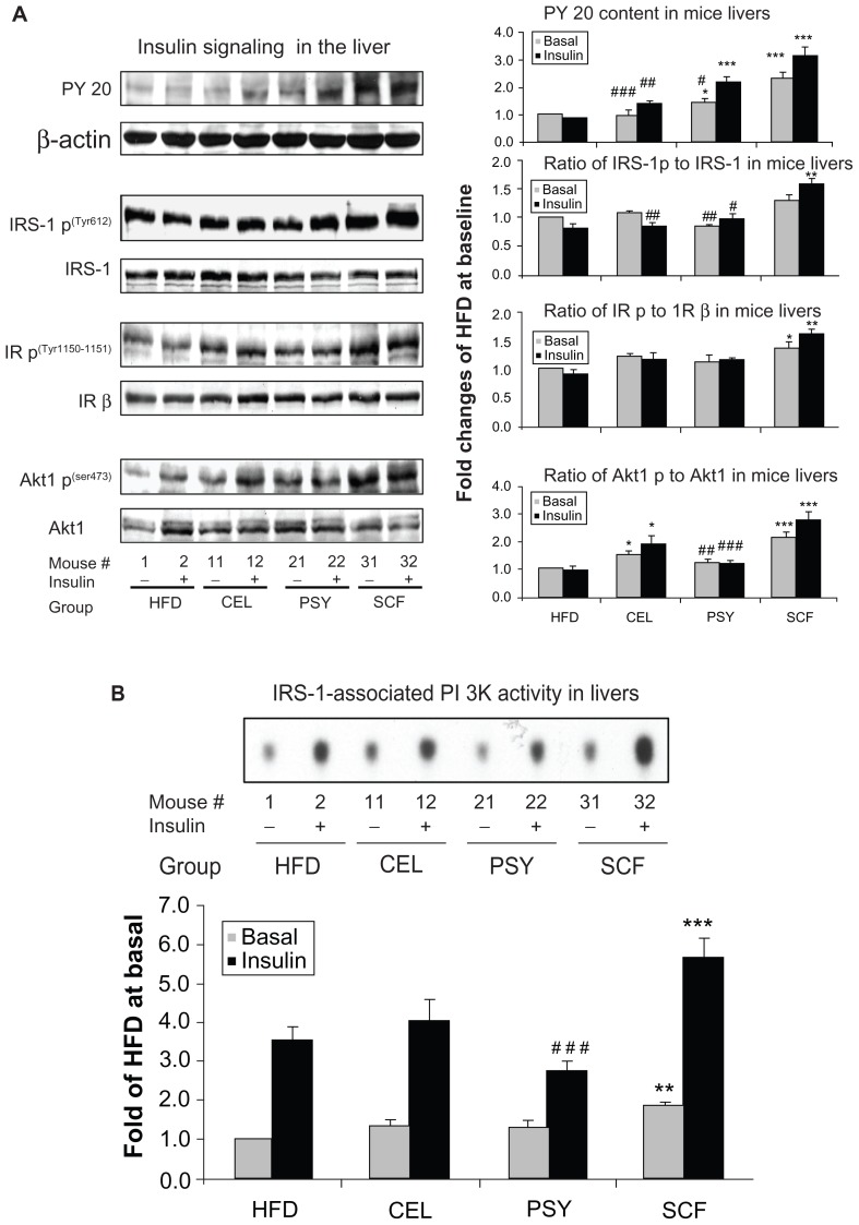
Figure 4.
Effects of dietary fibers on insulin signaling protein abundance and phosphatidylinositol 3 kinase (PI 3K) activity in mice livers. Liver lysates were subjected to sodium dodecyl sulfate polyacrylamide gel electrophoresis and insulin signaling pathway proteins were detected with corresponding specific antibodies as shown in the figures. (A) shows that phosphorylation of antiphosphotyrosine 20 (PY 20), insulin receptor substrate (IRS) 1, insulin receptor beta (IR β), and Akt1 were normalized by their corresponding protein contents and PY 20 was normalized by β-actin. (B) Hepatic IRS-1-associated PI 3K activity in mice. Liver lysates from basal and insulin-stimulated mice were immunoprecipitated with IRS-1 antibody and protein A agarose. The immune complexes were incubated with reaction buffer containing [γ-32P]adenosine 5′-triphosphate, MgCl2, MnCl2, and phosphatidylinositol for 20 minutes. Autoradiograph was performed after thin-layer chromatography.
Notes: Data presented as mean ± standard error of the mean (n = 9/group). *P < 0.05; **P < 0.01; ***P < 0.001, dietary fiber-supplemented group vs HFD-only group; #P < 0.05; ##P < 0.01; ###P < 0.001; CEL or PSY vs SCF group.
Abbreviations: CEL, cellulose; HFD, high-fat diet; PSY, psyllium; SCF, sugar cane fiber.

