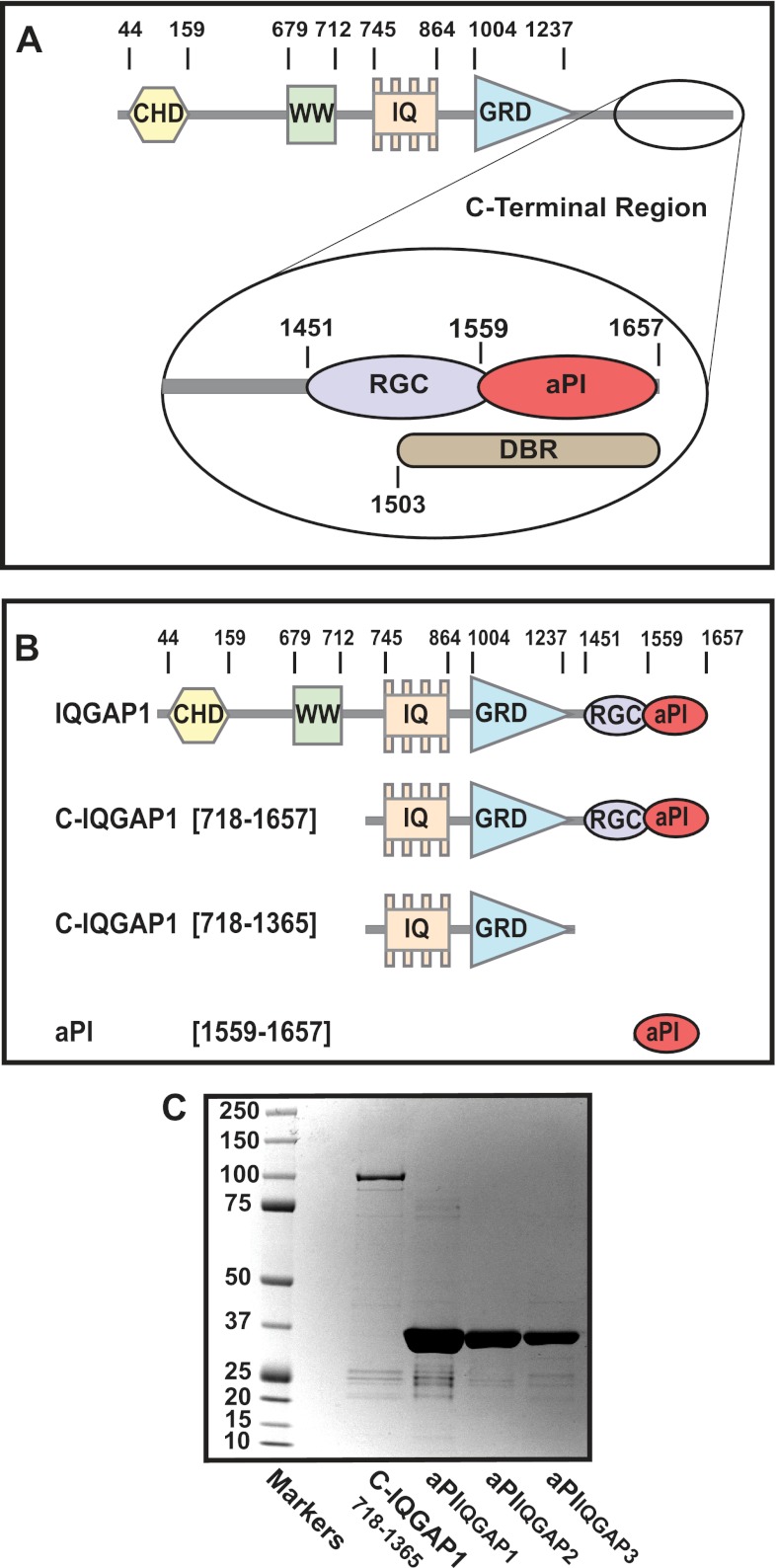
FIGURE 1.
The architecture of the IQGAP1 protein shows the location of the aPI binding module. A, the domain architecture of IQGAP1. The linear sequence of the established, functional domains within IQGAP1 is shown. These include the calponin homology domain (CHD), the proline-rich WW domain (WW), the repeat IQ motifs (IQ), and the RasGAP-related domain (GRD). The expanded view of the C terminus shows the RasGAP C terminus (RGC), Dia1-binding region (DBR), and the atypical PI binding module (aPI). B, the truncated IQGAP1 constructs examined for 3-PI binding. The central panel shows a schematic representation relative to full-length IQGAP1 of the three GST-tagged, truncated IQGAP1 constructs, C-IQGAP1(718–1657), C-IQGAP1(718–1365), and C-IQGAP1(1559–1657) (aPIIQGAP1) examined for 3-PI binding by surface plasmon resonance. C, the purity of IQGAP1 constructs. The purity of each construct determined by SDS-PAGE and colloidal blue staining is indicated. The results shown are representative of at least two separate preparations of each construct. The identity of the bands indicated was confirmed by mass spectrometry as described under ”Experimental Procedures.“

