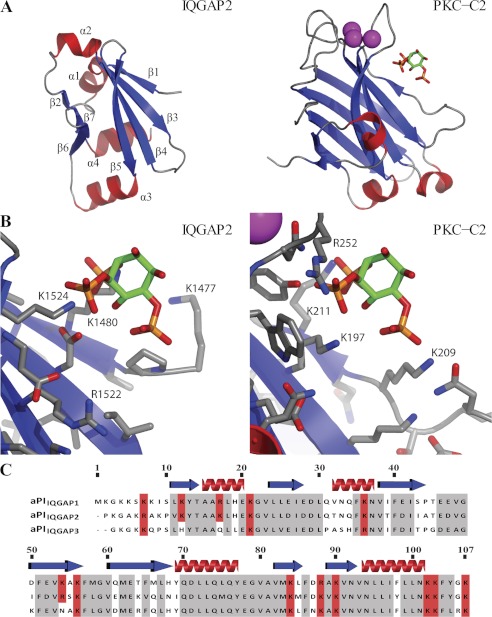
FIGURE 7.
The three-dimensional structure of the IQGAP2 C-terminal or aPIIQGAP2 domain. A and B, comparison of the IQGAP2 C-terminal domain structure (PDB code 4EZA) to that of the C2 domain of protein kinase Cα (PDB code 3GPE and (46)) in complex with the headgroup of PtdIns(4,5)P2. Structures are shown with the same viewing matrix after superposition. Secondary structure is shown with red helices and blue strands. Calcium ions are shown as magenta spheres. The PtdIns(4,5)P2 headgroup is shown as sticks. The position of the PtdIns(4,5)P2 headgroup in the protein kinase Cα C2 domain complex structure is also shown together with the IQGAP2 C-terminal domain in panel B to facilitate comparison of interacting side chains. C, a sequence alignment of the aPI domains from the IQGAP protein family (aPIIQGAP1, 1551–1657; aPIIQGAP2, 1470–1575; aPIIQGAP3, 1527–1631) reveals a high level of homology shown in gray. Basic amino acids conserved in aPIIQGAP1 and aPIIQGAP2 are shown in red. The absence of two of these, numbered 12 and 54 (corresponding to Lys-1562 and Lys-1604 in aPIIQGAP1 and Lys-1480 and Arg-1522 in aPIIQGAP2) from aPIIQGAP3, coupled to their location in the proposed binding pocket shown in panel B, suggests a possible role in ligand recognition.

