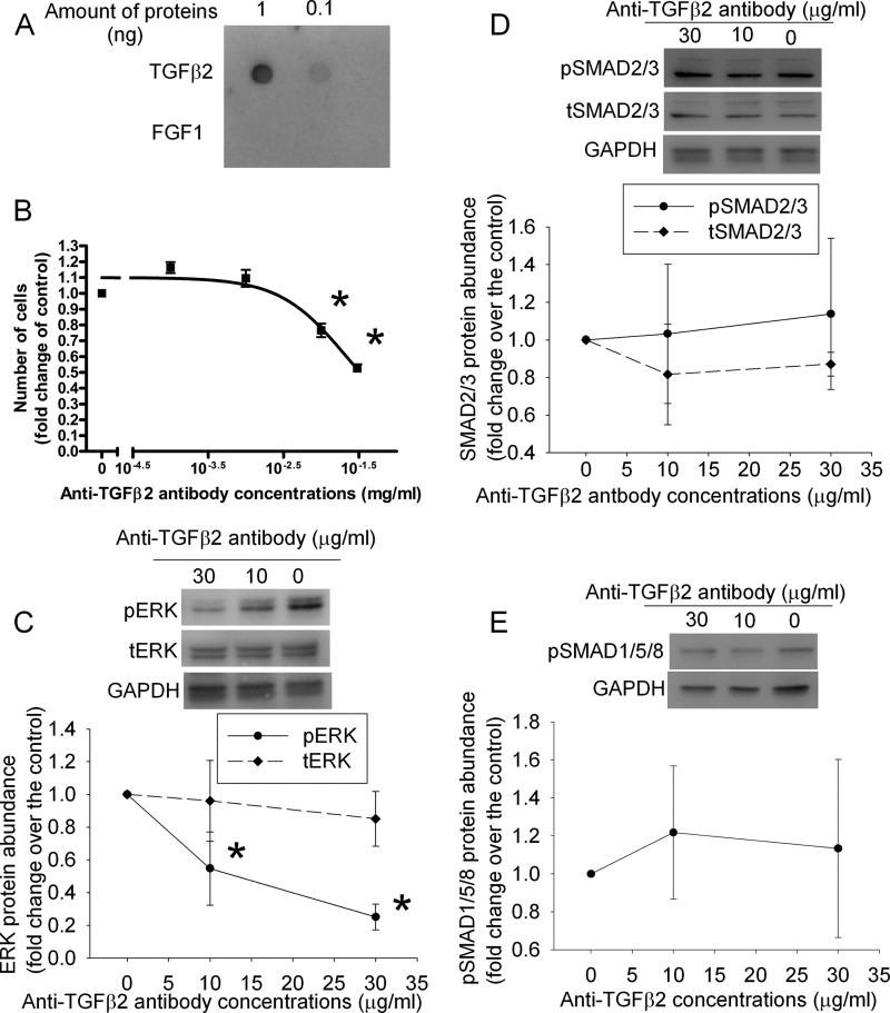
FIGURE 3.
Inhibition of cell proliferation and phospho-ERK expression by an anti-TGFβ2 antibody in the SH-SY5Y cells. A, a dot blot produced by loading 0.1 or 1 ng of TGFβ2 or FGF1 on the membrane and visualizing the proteins by an anti-TGFβ2 antibody. B, cells were cultured in medium without fetal bovine serum for 24 h and then with 10% fetal bovine serum in the presence of various concentrations of an anti-TGFβ2 antibody for 2 days. Cell numbers then were measured and normalized by those at the beginning of fetal bovine serum stimulation. C–E, cells treated as described for panel B were harvested for Western blotting of phospho-ERK (pERK), total ERK (tERK), phospho-SMAD2/3 (pSMAD2/3), total SMAD2/3 (tSMAD2/3), and phospho-SMAD1/5/8 (pSMAD1/5/8). The ERK and SMAD Western blot results were normalized by those of glyceraldehyde-3-phosphate dehydrogenase (GAPDH). The results from cells incubated with the anti-TGFβ2 antibody were then normalized by those from control cells. Results are mean ± S.D. (n = 3–4). *, p < 0.05 compared with the corresponding results without the anti-TGFβ2 antibody.

