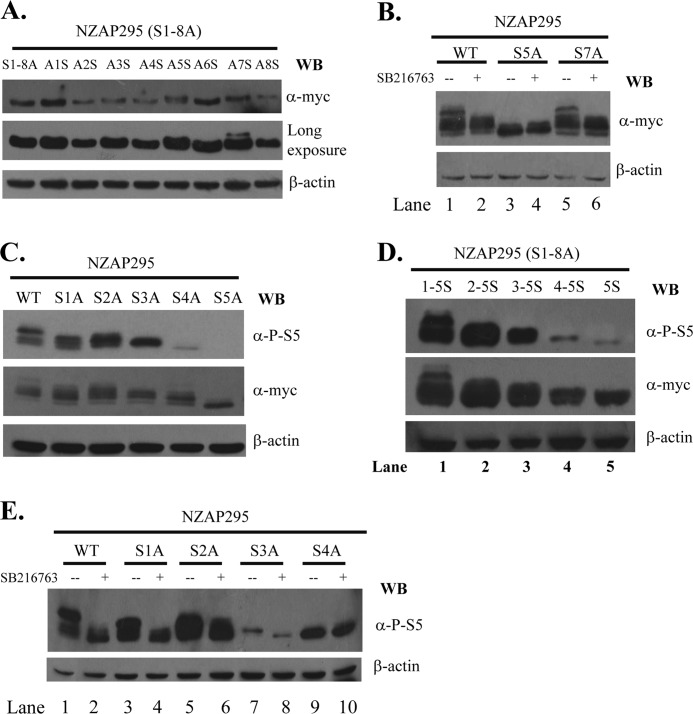
FIGURE 3.
Mapping serine residues phosphorylated by GSK3β. A, C, and D, the proteins indicated were transiently expressed in HEK293 cells, subjected to SDS-PAGE, and detected by Western blotting (WB) with the antibodies indicated. B and E, a plasmid expressing the proteins indicated was transfected into HEK293 cells. At 6 h post-transfection, the cells were mock-treated (−) or treated with SB216763 (+). Cells were lysed at 48 h post-transfection, and the lysates were analyzed by Western blotting with the antibodies indicated.

