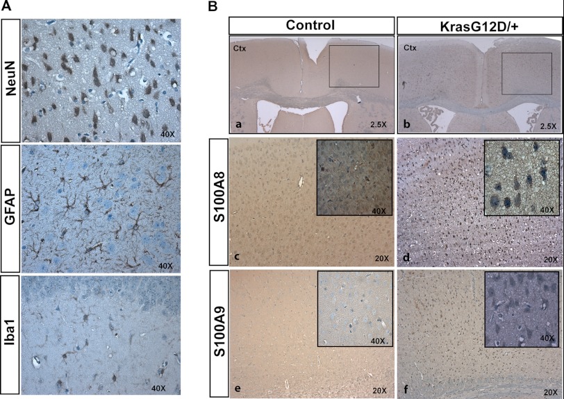
FIGURE 4.
S100A8 and S100A9 are overexpressed in Kras G12D/+ neurons. Cortices were isolated from 12-week-old control and CamKII-Cre; LSL Kras G12D/+ (Kras G12D/+) mice (n = 3). Serial coronal sections were cut at 5 μm thickness (paraffin) and analyzed by NeuN, GFAP, Iba1, S100A8, and S100A9 immunohistochemical staining. A, representative images of NeuN, GFAP, and Iba1 immunoreactivities to show the morphologies of neurons, astrocytes, and microglia, respectively, in Kras G12D/+ cortex. B, immunohistochemical analysis of S100A8 and S100A9 in control and Kras G12D/+ cortex. Panels c and d, high magnification views of the boxed area in panels a and b. The insets in panels c–f show high magnification views of cells overexpressing S100A8 in panels c and d or S100A9 in panels e and f. Morphologically, S100A8- or S100A9-positive cells resemble neurons. Ctx, cerebral cortex.

