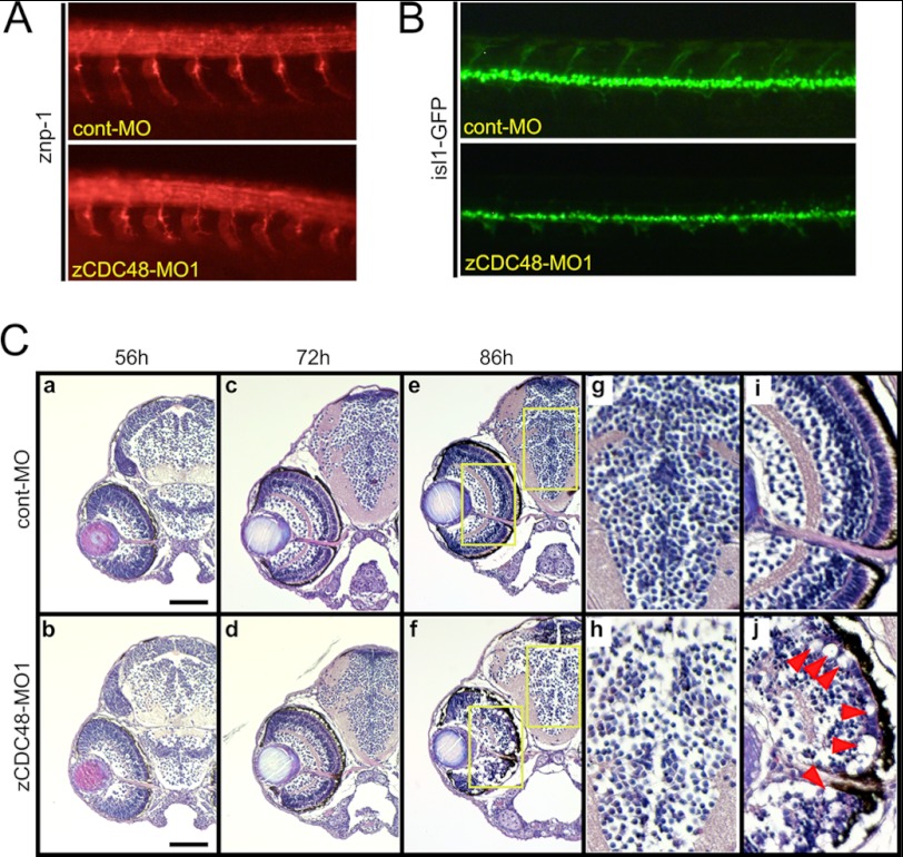
FIGURE 2.
Spinal motor axon defects and decreased numbers of neural cells in the eyes and brains of CDC48-deficient embryos. A, primary and secondary motor axons in zebrafish embryos were visualized by immunostaining with the anti-znp-1 (anti-synaptotagmin 2) antibody. By 36 hpf, secondary motor axons had extended into the ventral muscle and fasciculate to form a nerve. The zCDC48-MO-injected embryos had truncated and branched axons, compared with those of the control. B, primary and secondary motor axons in isl1-GFP-transgenic zebrafish are visualized. C, H&E staining revealed almost no changes in histology and normal development of the eye and brain until 72 hpf. After 86 hpf, the number of cells in the diencephalon decreased significantly; e is magnified to g (diencephalon) and i (eye), and f are magnified to h (diencephalon) and j (eye). The structures of the inner plexiform layer and ganglion cell layer are almost decomposed, the rods and cones have disappeared (j, red arrowhead), and the optic nerve has narrowed (j).

