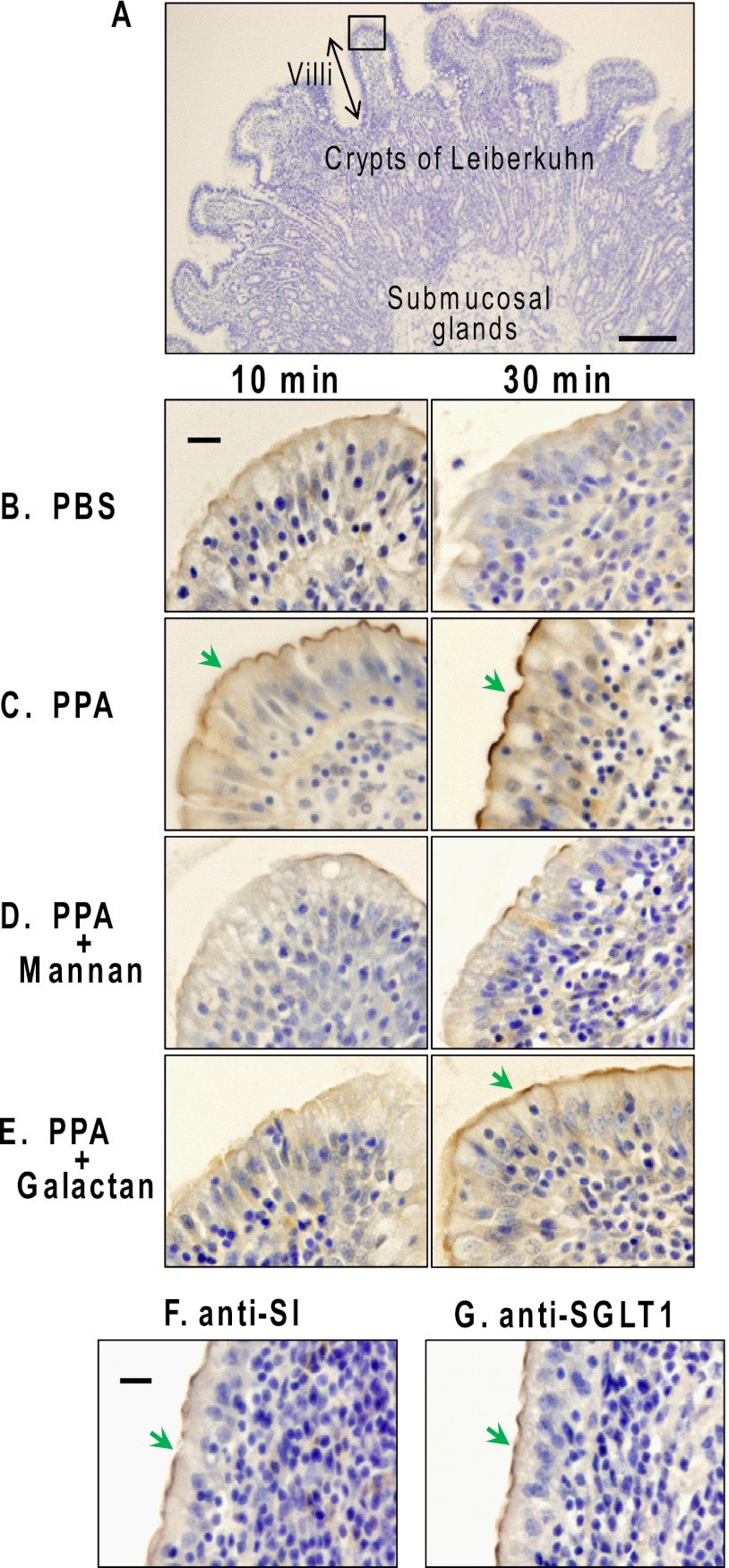
FIGURE 1.
Immunohistochemical staining of PPA in porcine duodenum. Porcine duodenum sections (A) from fasted animals were incubated with 20 mm PBS (B) or various PPA solutions (C–E) containing 1 mm phenylmethylsulfonyl fluoride at 4 °C for 10 and 30 min and then fixed and paraffin-embedded as described under “Experimental Procedures.” The paraffin sections were immunostained with rabbit anti-α-amylase IgGs-HRP (B–E). The color was developed with DAB/H2O2 and then counterstained with hematoxylin. Green arrows indicate positive staining on the apical surface (A) the luminal side of the duodenum. The tip of a villus surrounded by a square in A is shown in B–G. Duodenum sections were incubated in 20 mm PBS (B), 10 μm PPA (C), 10 μm PPA containing mannans (D), or galactans (E). Untreated porcine duodenum sections were detected with rabbit anti-SI IgGs-HRP (F) and SGLT1 (G). Scale bar in A, 100 μm; scale bars in B and F, 20 μm. Similar experiments were repeated three times.

