
FIGURE 7.
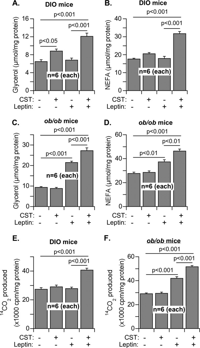
Lipolysis and fatty acid oxidation in the adipose tissue explants of DIO and ob/ob mice after treatment with saline or CST for 16 days. A–D, explants were incubated with saline or leptin (1 μm) for 3 h, and the concentrations of glycerol (A and C) and NEFA (B and D) released into the media from DIO (A and B) and ob/ob (C and D) explants were determined as a measure of lipolysis. E and F, homogenates of the explants from DIO (E) and ob/ob (F) were used to determine their capacity for oxidation of [U-14C]palmitate in response to the treatment with saline, CST, leptin, and CST + leptin. The 14CO2 released was captured and counted as the measure of fatty acid oxidation.