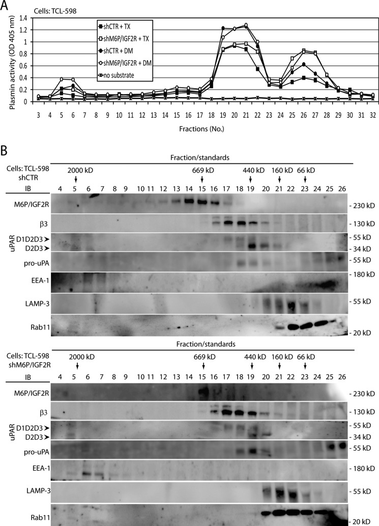
FIGURE 3.
Analysis of M6P-IGF2R complexes activating Plg in vitro. A, cell lysates derived from control (shCTR)- and M6P-IGF2R-silenced (shM6P-IGF2R) TCL-598 cells were prepared as described under “Experimental Procedures” by solubilization with either detergent Triton X-100 (TX) or detergent N-dodecyl-d-maltoside (DM). The samples were loaded onto a Superose 6 column, and individual fractions were collected. The fraction samples were transferred into wells of a 96-well plastic plate and incubated at 37 °C with human Glu-Plg (50 nmol/liter). Plasmin activities were analyzed by adding the chromogenic plasmin substrate (S-2251; 0.8 mmol/liter). The absorbance change at 405 nm was monitored after 4 h by using a 96-well plate reader. B, the fraction samples from A were analyzed by SDS-PAGE followed by immunoblotting (IB) with mAbs specific to M6P-IGF2R (mAb MEM-238), β3 integrin (mAb VIPL-2), uPAR (mAb H2), pro-uPA (mAb U5), Rab11 (polyclonal), LAMP-3 (mAb MEM-259), and EEA1 (mAb C45B10). Chemiluminescence was used for visualization. D1D2D3 points to the band corresponding to the full-length uPAR, and D2D3 points to the band corresponding to the truncated form. Results are representative for three independent experiments. Molecular mass standards used in A are specified under “Experimental Procedures.”

