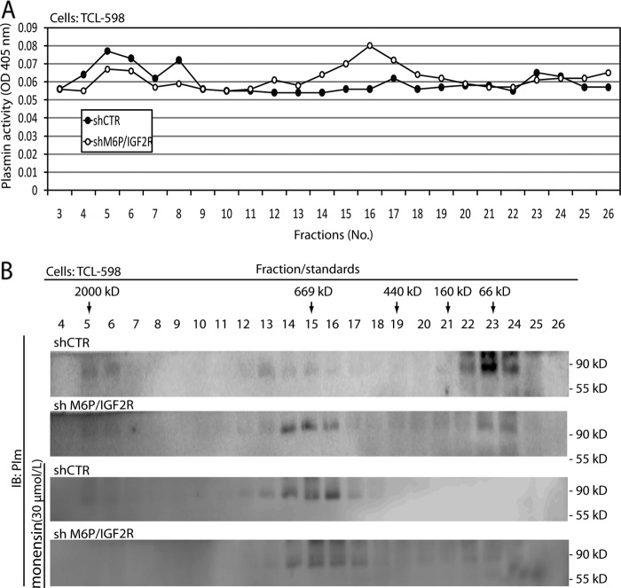
FIGURE 4.
Analysis of M6P-IGF2R complexes activating Plg in vivo. A, control (shCTR)- and M6P-IGF2R-silenced (shM6P-IGF2R) TCL-598 cells were incubated for 30 min with human Glu-Plg (50 nmol/liter) on ice followed by an additional 60-min incubation without Plg at 37 °C. Then the cells were solubilized in detergent Triton X-100, and the cell lysates were subjected to size exclusion using a Superose 6 column. Individual fractions were assayed for plasmin activity in the presence of the chromogenic plasmin substrate S-2251 (0.8 mmol/liter) as described in Fig. 3A. The absorbance change at 405 nm was monitored after 18 h by using a 96-well plate reader. B, the fraction samples were analyzed by SDS-PAGE followed by immunoblotting (IB) with rabbit polyclonal anti-plasmin Ab. In some settings the cells were preincubated with monensin (30 μmol/liter).

