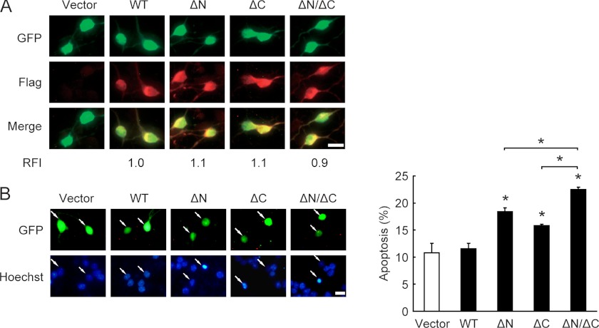
FIGURE 6.
Truncated forms of GSK-3β promote neuronal death. A, DIV5 CGNs were transfected with FLAG-tagged GSK-3β WT, ΔN, ΔC, or ΔN/ΔC along with GFP for 36 h, and the fluorescence intensity was quantified by immunostaining with the FLAG antibody. The relative fluorescence intensity (RFI) of GSK-3β WT-transfected cells was set to 1.0. Scale bar, 10 μm. B, apoptosis was determined according to the percentage of GFP-positive cells showing pyknotic nuclei (stained by Hoechst 33258). Representative images are shown (left panel). Data represent the mean ± S.E. of three experiments. *, p < 0.05. Scale bar, 10 μm.

