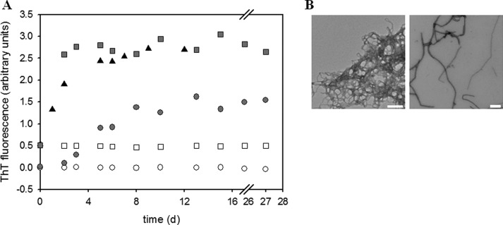
FIGURE 9.
Fibril formation kinetics of truncated PABPN1 variants. A, fibril formation of SUMO-ΔN114 (triangles), ΔAla-N147 (open symbols), and (+7)Ala-N147 (filled symbols). Samples seeded with 5% (w/w) seeds from N-(+7)Ala (21) are indicated by squares, unseeded samples by circles. B, electron micrographs of fibrillar SUMO-ΔN114 (left) and of (+7)Ala-N147 (right).

