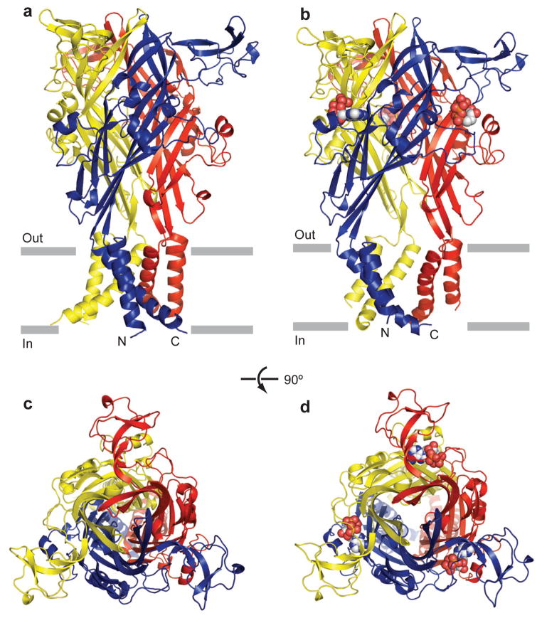
Figure 1. The architectures of zebrafish P2X4.
a, b, zebrafish ΔP2X4-B2 (a) and ΔP2X4-C (b) trimer structures viewed parallel to the membrane. Each subunit is shown in a different color. ATP is shown in sphere representation. c, d, zebrafish ΔP2X4-B2(c) and ΔP2X4-C (d) trimer structures viewed from the extracellular side.

