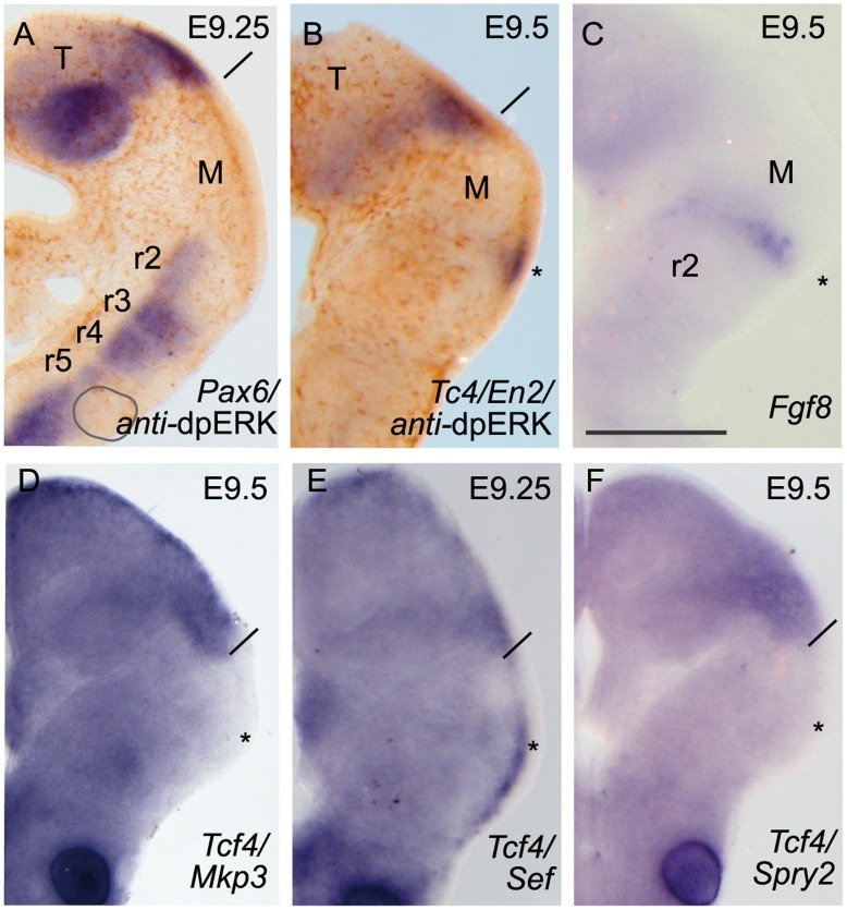
Figure 2. Low threshold of FGF8 protein levels disrupts ERK1/2 phosphorylation patterns.
.DpERK immunodetection was absent in the isthmic domain using ONTCs severe hypomorphic mouse mutant (A, B; Fgf8 neo/null [45]). Yet a small tip of En2 (B) positive expression was visible at the most dorsal parts, probably by the maintenance of Fgf8 (C) expression. Under these mutant conditions, none of the FGF8 signal negative modulators Mkp3 (D), Sef (E) Sprouty2 (F) were observed at IsO. Asterisks indicate the position of abolished isthmic region and solid line the boundary between diencephalon/mesencephalon. Scale bar in C is 0,5 mm for all images.

