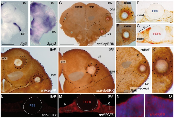
Figure 4. Bafilomycin A1 (BAF) treatment demonstrates the polarization of ERK activity by FGF8 signal activity along the neural tube.
A-C) BAF amplifies ectopic ERK1/2 activity-related FGF8 induction revealing a clear polarized distribution of ERK1/2 activity around FGF8 soaked beads depending of the implanted bead; rostral to IsO (C,D,G), or caudal to IsO (C,E). Nonetheles, isthmic organizer morphogenetic activity seems unaffected for Fgf8 (A) and negative regulator Sprouty 2 (B) expressions. Note that PBS bead implantation in control side (blue asterisk in C and F) did not show any ectopic induction. Two hours after bead implantation a clear amplified and almost non-homogeneous ERK1/2 activity was detected rostrally in the mesencephalon (rostral to the IsO), which was detected caudally when bead was placed in hindbrain (caudal to the IsO) territories (E). In telencephalic vesicles, caudal to the anr (H) the polarity of ERK activation was reversed. This polarized dpERK detection around the bead is lost at the zli (zona limitans intrathalamica) region (I). Similar symmetric ERK-related FGF8 signal found in zli was seen when placing a FGF8 bead in the midbrain of Fgf8 hypomorphic mice (J,K). Importantly FGF8b protein distribution (M) was observed apparently in equal intensity and range at rostral (N) and caudal (O) sides of the bead (for comparison with PBS bead in panel L). Scale bars are 0,5 mm in A, B, C, H, I, 200 µm in D, E, J, 100 µm in F, G, K, L, M, and 50 µm in N, O.

