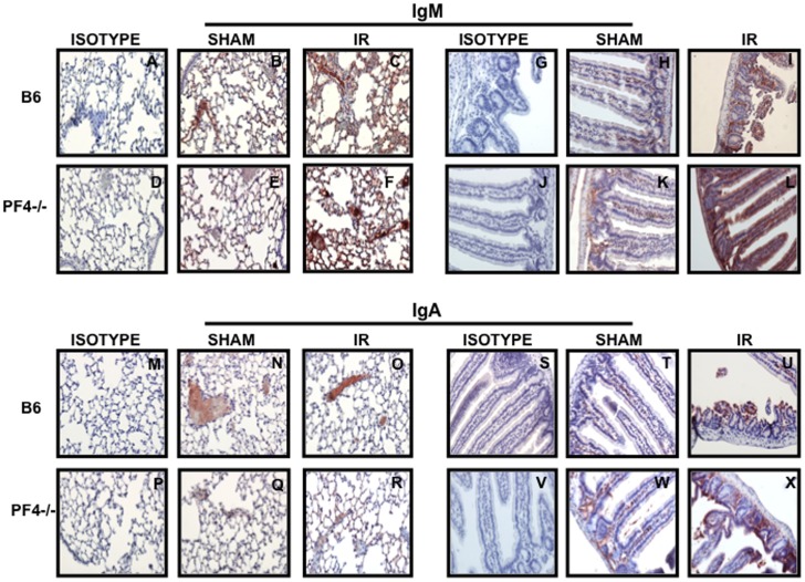
Figure 5. Tissue damage in PF4-/- mice is not associated with immunoglobulin (Ig) deposition.
Tissue sections of lung and intestine from B6 and PF4-/- mice after 30 minutes of mesenteric ischemia and 3 hrs of reperfusion were stained for IgM (A-L, red) and IgA (M-X, red) and counterstained with hematoxylin (blue). Images are representative of 3–4 mice per group. Red: Positive Staining.

