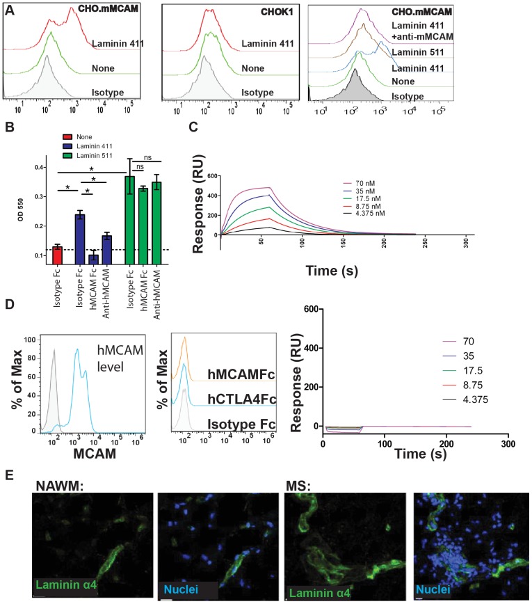
Figure 5. Laminin 411 functions as a ligand for MCAM.
(A) CHO cells transfected with hMCAM were incubated with recombinant laminin 411. Laminin binding to the surface of the cells was detected with an anti-laminin antibody (left panel), while no binding was detected to CHO cells lacking MCAM expression (center panel). Recombinant laminin 511 did not bind to MCAM expressing CHO cells, and binding of laminin 411 was specifically inhibited by preincubation of the cells with anti-hMCAM (clone 17, right panel). (B) Human CD4+CD45RO+ memory T cells were purified and incubated with TCR stimulation in the presence of TGFβ and IL1β to induce hMCAM expression (approximately 50% expressed hMCAM, similar to Figure 2A). Cells were incubated in plates coated with either laminin 411 or laminin 511 in the presence of either hMCAM-Fc or anti-hMCAM and specific binding of the cells to the laminin was determined. * indicates p<0.05. (C) Binding of recombinant human laminin 411 to immobilized hMCAM-Fc protein was detected by Biacore with a Kd of approximately 27 nM. Data is representative of three individual experiments. (D) mMCAM is expressed at high levels in nearly all CHO cells transfected with the mMCAM gene, and sorted for high expressors (blue histogram compared to shaded isotype control). However, soluble mMCAM protein does not bind to these cells at levels higher than either human IgG control or irrelevant Fc tagged protein, CTLA-4. Furthermore, no specific binding of mMCAM-His to mMCAM-Fc was detected by Biacore. (E) Laminin α4 was detected in both human MS lesions and normal appearing white matter.

