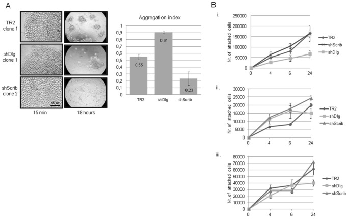
Figure 5. hScrib depleted cells exhibit reduced levels of cell-cell contact.
Panel A. Cells were suspended from a plastic tissue culture dish lid in a drop of medium and incubated overnight. The cells were photographed after 15 mins and after 18 h incubation in suspension, where a single cell suspension can be seen at the 15 mins time point and aggregates of cells at the 18 h time point. The graph shows the aggregation index calculated from at least three independent experiments using two independent control, hDlg1 and hScrib depleted cell lines. Error bars are also shown. Panel B. Cells were plated on plastic tissue culture dishes that were either untreated (i), coated with fibronectin (ii) or coated with collagen (iii) and the attached cells counted at different time points. The graphs show the results from at least three independent assays using three independent control, hDlg1 and hScrib depleted cell lines and the standard deviations are shown.

