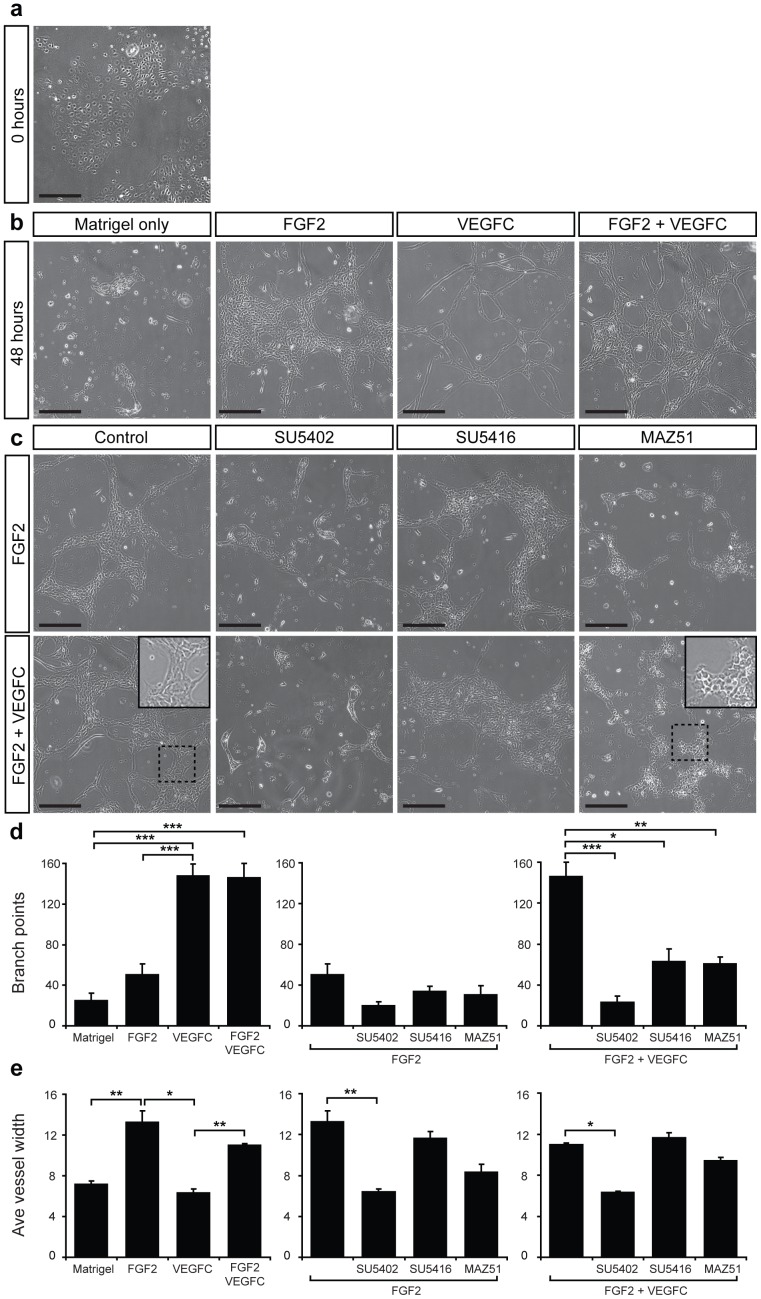
Figure 5. FGF2 and VEGF-C promote tube formation of primary mouse LEC.
(a) Primary LEC were cultured for 24 h and imaged immediately following the addition of Matrigel. (b) Primary LEC were cultured for 24 h followed by addition of Matrigel alone or Matrigel containing FGF2 (10 ng ml−1), VEGF-C (200 ng ml−1) or a combination of FGF2 and VEGF-C. Images were captured after a further 48 hours. (c) Primary LEC were cultured for 24 h followed by addition of Matrigel containing FGF2 (10 ng ml−1) or a combination of FGF2 (10 ng ml−1) and VEGF-C (200 ng ml−1) and tyrosine kinase inhibitors SU5402 (10 µM, FGFR1), SU5416 (5 µM, VEGFR-2) or MAZ51 (5 µM, VEGFR-3). Three replicates of each treatment were performed and images are representative of at least three independent cell isolations. Inset panels in (c) illustrate magnified views of boxed regions. Scale bars represent 250 µm. Quantification of average vessel diameter (d) using Lymphatic Vessel Analysis Protocol (LVAP) [28] and ImageJ [29] software and branch points per well (e) using AngioTool software [30], for each treatment indicated. Data show mean ± s.e.m. and are derived from 2 independent cell isolations, each prepared from multiple litters of embryos, and 3 replicates of each treatment (n = 6). *P<0.05, **P<0.01, ***P<0.001.

