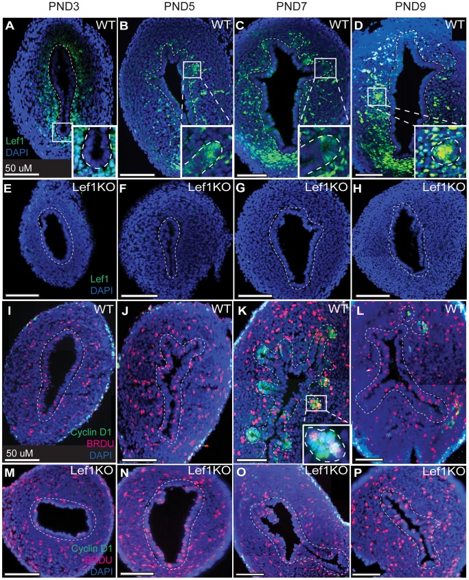
Figure 4. Lef1 and cyclin D1 are expressed during gland development in WT but not Lef1 KO mouse uteri.
(A–P) Lef1 and/or cyclin D1/BrdU expression in the developing uterus on post-natal days (PNDs) 3 (A, E, I, M), 5 (B, F, J, N), 7 (C, G, K, O) and 9 (D, H, L, P). (A–D) Photomicrographs of Lef1 expression in wild-type uteri. Expression is present in both mesenchymal and epithelial tissue throughout this period, and is visible in the glands as they begin to form on PND 5 (panel B and inset). (E–H) Photomicrographs of Lef1 expression in Lef1 KO mice. In addition to lacking Lef1 expression, these uteri fail to form endometrial glands. (I–L) Photomicrographs of cyclin D1 expression and BrdU incorporation in wild-type uteri. Both are visible in developing endometrial glands beginning on PND 7 (panel K and inset). (M–P) Photomicrographs of cyclin D1 expression and BrDU incorporation in the uteri of Lef1 KO mice. In addition to an absence of formed glands, these uteri lack cyclin D1 expression despite the fact that cell proliferation is active, as indicated by BrdU incorporation. Dashed lines mark the border between the mesenchymal and epithelial cell layers.

