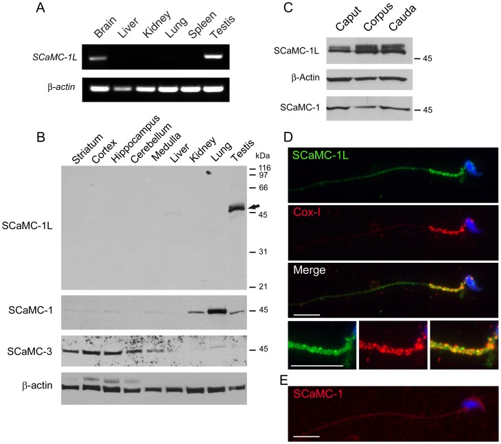
Figure 2. SCaMC-1L is expressed in testis and male germ cells.
(A) RT-PCR analysis of SCaMC-1L expression in mouse tissues. Equivalent aliquots of cDNAs derived from the indicated tissues were used as templates. Amplification of β-actin was used as an internal control. The results obtained indicate that SCaMC-1L is expressed preferentially in testis and, at lower levels, in brain. (B) Expression of SCaMC-1L protein in adult mouse tissues. 10 µg of total protein extracts from the indicated tissues were analyzed by western blot using a specific anti-SCaMC-1L antibody. A single band of the expected size, around 50 kDa, was exclusively detected in testis, marked with an arrow. β-actin levels are shown as loading control. A parallel blot was incubated with antibodies against SCaMC-1 which detected a single band of 45 kDa, and then with antibodies against SCaMC-3 which detected a band of of about 48 kDa. The specific distribution patterns of the labelled bands in mouse tissues rule out any significant crossreactivity among paralogs. (C) 5 µg of total proteins from cauda, corpus and caput spermatids were analyzed by western blot with anti-SCaMC-1L antibody. Membranes were re-probed with β-actin antibody as loading control and anti-SCaMC-1. Equivalent SCaMC-1L levels are found in spermatids from different regions of epididymis. (D) SCaMC-1L-staining is detected in the midpiece of epididymal spermatids. SCaMC-1L was detected using an affinity-purified SCaMC-1L antibody and visualized with a FITC-conjugated secondary antibody, mitochondria were stained with an anti-COX-I monoclonal antibody and visualized with a Cy3-conjugated secondary antibody, nuclei were stained with Hoechst and triple merged panel is also shown. Merge panel shows the co-localization of SCaMC-1L and COX-I staining in the mitochondrial sheath of midpiece. Enlarged images of this region are shown at bottom. (E) SCaMC-1 is not detected in mature spermatids. SCaMC-1 staining was performed using anti-SCaMC-1 antibody at dilution 1∶200 and visualized Alexa Fluor 555 anti-rabbit as secondary antibody, nuclei were stained with Hoechst. Scale bars; 10 µm.

