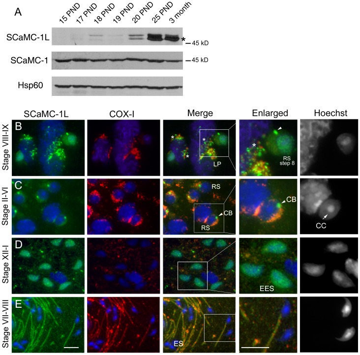
Figure 3. Immunohistochemical localization of SCaMC-1L protein in mouse testis.
(A) Analysis of SCaMC-1L expression during spermatogenesis. SCaMC-1L levels were determined in mitochondrial-enriched extracts prepared from mouse testis at different post-natal days (PND), between 15–25 days and 3-month-old, by western blot. The levels of the mitochondrial proteins SCaMC-1 and hsp60 were used as loading controls. (B–E) Detection of SCaMC-1L in squash preparations of seminiferous tubules by immunofluorescence assays. The stages of seminiferous segments are indicated. SCaMC-1L and COX-I detection were performed as described in Figure 2. SCaMC-1L is detected in granules close to mitochondria in late pachytene spermatocytes (LP) (B), and round spermatids (RS) (B, C), identified by the heterochromatic chromocenter (CC) at the nucleus (C, indicated by an arrow). In elongating spermatids (EES) a diffuse SCaMC-1L staining is detected in the nucleus (D), in elongated spermatid (ES) SCaMC-1L is found at the midpiece matching with COX-I signals (E). Enlarged images of marked insets for merged panels and corresponding Hoechst staining are shown. Scale bars; 10 µm.

