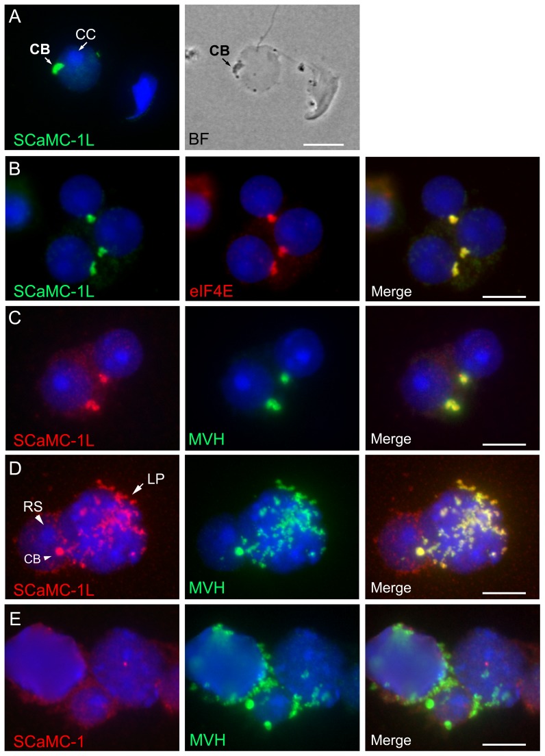
Figure 5. Localization of SCaMC-1L in chomatoid body.
Drying-down slides were labeled with anti-SCaMC-1L antibody and its localization in chromatoid body (CB) of round spermatids (RS) was confirmed by parallel phase contrast microscopy (A) and by co-staining with the specific markers for CB, eIF4E (B) and MHV (C, D). CBs, identified as eIF4E and MVH protein-positive structures, appear strongly stained for SCaMC-1L. In late pachytene spermatocytes (LP) SCaMC-1L signals co-localizes entirely with cytosolic granules MVH-positive (D). RS were identified by the heterochromatic chromocenter (CC) at the nucleus (indicated by arrow in A). (E) Absence of co-localization of SCaMC-1 and MVH signals in drying-down slides. Scale bars; A, 5 µM; B–E; 10 µM.

