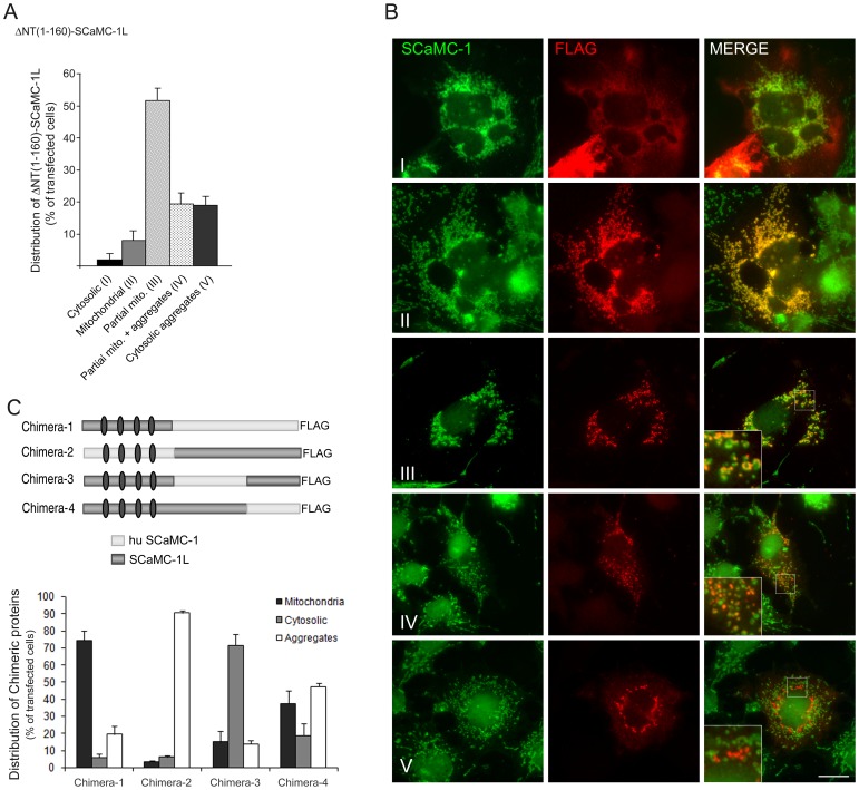
Figure 8. Both N-extension and C-half MC regions of SCaMC-1L are involved in its intracellular distribution.
(A) The N-terminal extension of SCaMC-1L hampers its import into mitochondria. COS-7 cells were transfected with a FLAG-tagged amino truncated SCaMC-1L protein, ΔNT(1–160)-SCaMC-1LFLAG, and 24–30 h later were fixed and co-stained with anti-FLAG (red) and anti-SCaMC-1 (green) antibodies. FLAG and SCaMC-1 images were taken under identical conditions and co-localization was evaluated in merged compositions. Five patterns were clearly identified (I–V). Most of ΔNT(1–160)-SCaMC-1L-positive cells show, at distinct degree, mitochondrial localization. These cells were sub-classified according to their co-localization degree with mitochondria (II–III) and the additional presence of SCaMC-1L aggregates (IV). Results are the mean ± SEM of three independent experiments. At least 50 cells were analyzed for each transfection assay. (B) Representative images of FLAG/SCaMC-1 double stained cells showing ΔNT(1–160)-SCaMC-1L-patterns I, II, III, IV and V merged panels are also shown. Insets magnification 300×; scale bar, 20 µM. (C) Scheme of the chimeric SCaMC-1L/SCaMC-1 proteins used. Relative positions of the EF-hand calcium-binding domains are marked by gray ovals. Quantification of the intracellular patterns observed for each protein (bottom) was performed as described in Figure 6. One hundred cells were counted for each transfection assay. Results are the mean ± SEM of three independent experiments.

