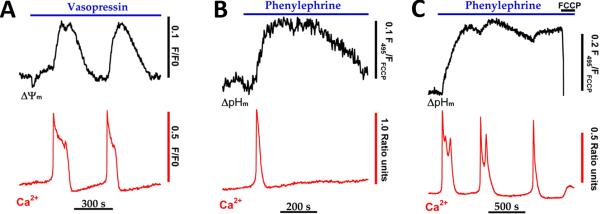
Fig. 3. Cytosolic calcium spikes stimulate a rise in mitochondrial proton motive force.
Hepatocytes isolated from chow-fed rats were loaded with fura2/AM and TMREE (A) or fluorescein diacetate (B-C) then treated with submaximal hormone concentrations. Hormone-evoked increases in Ca2+ and mitochondrial membrane potential (ΔΨm) or Ca2+ and mitochondrial pH gradients (ΔpHm) were monitored simultaneously as described [17, 38]. The protonophore, FCCP (5 μM), was added in C to completely collapse mitochondrial PMF. The data in panel A are reproduced from [38] with permission.

