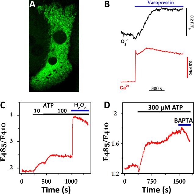
Fig. 4. Hormone-evoked increases in the rate of mitochondrial ROS formation.
(A) Maximal intensity projection of two adjacent hepatocytes expressing mitochondrial-targeted cpYFP, a superoxide-sensitive fluorescent protein. (B) Simultaneous measurement of cytosolic Ca2+ and mitochondrial superoxide (O2•-) increases evoked by maximal vasopressin concentrations (100 nM). Increases in Ca2+ were monitored with fura-2, while 485nm excitation of cpYFP was used to follow O2•- responses. Note: fura-2 overlaps with the 410nm portion of the cpYFP spectrum. (C-D) High levels of extracellular ATP evoke sustained increases in the production of mitochondrial H2O2. Production of H2O2 was monitored with mitochondrial targeted Hyper™ and alternating excitation at 485nm and 410nm. Exogenous H2O2 (100 μM) was added at the end of the run to maximally oxidize the biosensor (C). In panel D, excess BAPTA free acid (2 mM, blue bar) was added to chelate extracellular Ca2+.

