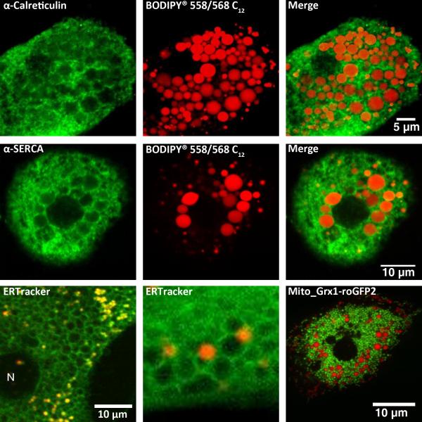
Fig 6. Lipid droplets.
Hepatocytes isolated from alcohol-fed rats (top row) or their pair-fed littermate controls (middle row) were maintained in primary culture for 4 hrs. Lipid droplets were labeled for 2-4 hrs with the red fluorescent fatty acid analogue Bodipy® 558/568 C12 (red pseudocolor). Cells from pair-fed controls (middle row) were also incubated with 300 μM oleic acid to induce lipid droplet formation. Cultures were fixed with 1% paraformaldehyde and then immunoreactivity for calreticulin or SERCA (kind gift from Dr. J. Lytton) was determined. Top row: calreticulin immunoreactivity in an alcoholic hepatocyte. Middle row: SERCA immunoreactivity in a lipid-loaded control hepatocyte.
Bottom row: Live hepatocytes from chow-fed rats were stained with 200 nM ER tracker (green pseudocolor) and Bodipy® 558/568 C12 (red pseudocolor), or transfected with a mitochondrial targeted glutathione biosensor Grx1-roGFP2 (kind gift from Dr. T. Dick, green pseudocolor) then incubated with the fatty acid analogue (bottom right most panel).

